Unfocused extracorporeal shockwave therapy diminishes bone loss in rats
Introduction:Extracorporeal Shockwave (ESW) therapy has shown to be effective in the treatment of non-unions and can enhance healing of fresh fractures(1). Enhanced proliferation and differentiation of osteoprogenitor cells have been shown in rats after a single ESW treatment(2). These studies used focused shockwaves; nowadays unfocused shockwaves can be generated, enabling treatment of larger areas without the need for anesthesia.Therefore extracorporeal shockwave therapy might be useful in the treatment of osteoporosis.
We evaluated if unfocused electro-hydraulically generated ESW can reduce bone loss and/or enhance fracture healing in ovariectomized and healthy rats.
Materials and Methods: 48 Female Wistar rats, 20 weeks of age were obtained. To induce bone depletion ovariectomy (OVX) was performed. ESW therapy (Dermagold, MTS-Europe GmbH, Konstanz, Germany) was given to the antero-lateral side of the right leg, the untreated left leg served as control. Two treatment regimes were used: a
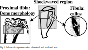
single treatment with 2000 pulses, 0.13 mJ/mm2, or two treatments with three weeks in between with 1000 pulses, 0.13 mJ/mm2.The legs were treated 3 (and 6) or 10 (and 13) weeks after OVX. Two days before the first ESW treatment a bilateral fibula osteotomy was performed to evaluate effects on fracture healing. A group with sham OVX and a fibula osteotomy was treated with 2000 pulses, to evaluate effects in healthy bone. Control groups to check for interactions between the osteotomy and ovariectomy were also evaluated. N=6 for all groups. Under gas anesthesia in-vivo microCT scans of the proximal tibia of the treated and untreated legs were made just before ESW treatment and 3, 6 and 10 weeks thereafter. In vivo microCT allows longitudinal follow-up of a single rat making it possible to evaluate dynamic bone changes in a highly detailed manner (voxelsize of 18 microns). Binary datasets of reconstructed images were made using a local threshold algorithm. A region of interest containing the proximal metaphysis was selected manually (length of 5,4mm), cortical and trabecular bone were automatically separated. Bone morphologic parameters of both were calculated (see Fig. 1). To evaluate the effect of ESW on fracture healing, a cylindrical region of interest 2mm in length was made around the osteotomy. A global threshold was used to determine the amount of mineralized matrix see (Fig. 1). All outcome measures were statistically evaluated using the Wilcoxin signed rank test.
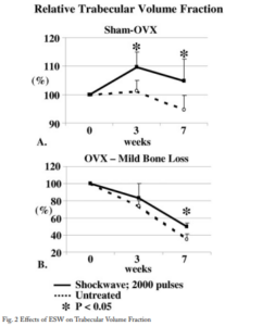
Results: ham-ovariectomized rats showed enhanced trabecular bone formation after shockwave therapy. Treated legs showed an increase of the trabecular volume fraction of 5% 7 weeks after treatment, whereas the non-treated legs showed a 5% decrease over the same period (p=0.03)(Fig. 2A). The cortical volume did not change significantly. Other bone morphologic parameters were not different in treated and untreated legs. The treated legs of rats suffering bone loss due to OVX that received shockwave therapy 3 weeks after OVX showed a slightly larger bone volume than the contralateral untreated legs, when they were treated with 2000 pulses (Fig. 2B). Cortical bone volume was not different between treated and untreated legs. No statistically significant effect of treatment was found in rats suffering bone loss due to OVX that received shockwave therapy after 10 weeks. There was no difference in mineralized callus formation in the fibula osteotomy between treated and non-treated legs in all treatment groups.
Discussion: A single treatment of 2000 pulses resulted in enhanced trabecular bone formation in sham-ovariectomized rats. The bone formation was only seen in the cancellous bone, which suggests that shockwave therapy influences bone remodeling and is therefore most effective in regions with a high remodeling rate. Cortical bone remodeling is almost absent in normal rats. The beneficial effect on trabecular volume fraction was also seen in rats that were shockwaved 3 weeks after OVX. However in rats that were treated 10 weeks after OVX no effect of shockwave was found. This suggests that ESW affects bone remodeling and causes effects in existing bone structures, but cannot induce de novo bone formation. Because bone structures are still seen in the marrow cavity of patients suffering severe osteoporosis, ESW might be able to induce bone formation in these patients.
van der Jagt OP, Schaden W, van Schie H et al.Orthopaedic Research Society. 2008; Poster No. 964